

Oxygen is used to heal wounds!
What is HBOT?
HBOT (Hyperbaric Oxygen Therapy) is merging as an advancement in treating diseases such as wounds, infections, Alzheimer's and infertility to help towards healing. This is such a great advancement that can help many people today and is likely to grow in these types of devices.
HBOT is widely being used especially with people who have diabetes due to the complications of poor circulation which in many cases leads to loses of limbs.

How does HBOT work?
The patient inhales the oxygen which stimulates the atmospheric pressing of being 33 feet below sea level and boosts the body's natural healing process. Recently this type of therapy has been used as an alternative for various conditions like infertility and Alzheimer's disease.
HBOT helps provide more oxygen to the body's tissues so they could work efficiently. Additional oxygen can enhance the healing of damaged tissue, boost tissue function and fight infection.
It is an exciting time for medicine and look forward to seeing this treatment heal and save many more patients.
References: http://www.counselheal.com/articles/30572/20170117/hyperbaric-oxygen-therapy-promising-new-way-to-treat-diseases.htm

Being inactive can give you asthma

Leading an inactive lifestyle can increase a person's risk of poluution related asthma according to scientists in the USA.
The scientists found that, after being exposed to ozone the inactive tests shouwed inflammation in the lungs – a symptom of asthma. Exposure to ozone impacted on the breathing rate of both groups.
Ground-level ozone is a type of pollution created by chemical reactions between the sun's rays and organic gases and oxides of nitrogen emitted by cars, power plants, industrial boilers, refineries, chemical plants and other sources.
The researchers argue that this finding could also be similar among humans, but further research is needed.
reference: http://www.europeanlung.org

Can meat be bad for asthma?
Experts say people should eat no more than 70g a day of red and processed meat for good health with processed meat possibly making asthma symptoms worse.

A study carried out in France 'journal Thorax' believe the preservative nitrite used in sausages, ham and salami may make these symptoms worse. The survey looked specifically at asthma symptoms - breathlessness, wheeze, chest tightness - and intake of cured meat: a single portion was two slices of ham, one sausage or two slices of salami.
"People who said they consumed more than four portions a week - eight slices of ham or four sausages, for example - had the biggest deterioration of their asthma by the end of the study."
The experts stress that their work cannot prove diet is definitely to blame. There are lots of factors in a person's life that can make their asthma worse.
references: bbcnews.co.uk

Stephen Hawking takes a breakth and visits the Vatican
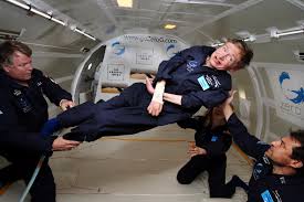
Stephen Hawking’s visit to the Vatican last month with Oxygen provided by our team. As stated online from the Catholic News Agency, Hawking is a member of the Pontifical Academy of Sciences – which includes 80 of the most brilliant scientists in the world – and he was in Vatican City for the group’s annual meeting. Hawking himself gave a talk on “The Origin of the Universe,” the topic that has earned him world renown.
His many publications include The Large Scale Structure of Spacetime with G F R Ellis, General Relativity: An Einstein Centenary Survey, with W Israel, and 300 Years of Gravity, with W Israel. Among the popular books Stephen Hawking has published are his best seller A Brief History of Time, Black Holes and Baby Universes and Other Essays, The Universe in a Nutshell, The Grand Design and My Brief History.
Professor Hawking has twelve honorary degrees. He was awarded the CBE in 1982, and was made a Companion of Honour in 1989. He is the recipient of many awards, medals and prizes, is a Fellow of The Royal Society and a Member of the US National Academy of Sciences.
Stephen was diagnosed with ALS, a form of Motor Neurone Disease, shortly after his 21st birthday. In spite of being wheelchair bound and dependent on a computerised voice system for communication Stephen Hawking continues to combine family life (he has three children and three grandchildren), and his research into theoretical physics together with an extensive programme of travel and public lectures.
During his conference at Casina Pio IV, Stephen Hawking paid homage to Msgr. George Lemaitre, president of the Pontifical Academy of Sciences from 1960 to 1966. Hawking said that Msgr. Lemaitre was the real father of the “Big Bang Theory,” thus dismissing the common belief that the father of the theory was the U.S. naturalized physicist George Gamow.
References

Christmas check list
 Christmas is a time for friends, family and a lot of preparing gifts, journeys, cards to send but most of all do not get too stressed and rushed and forget to look after yourself or your family's health over the festive period.
Plan ahead:
- Order any repeat prescriptions you may need especially if you are going away for the festive season.
- Check that you have enough oxygen supply for the Christmas period.
- If you are going away make sure you make arrangements for any oxygen that you need.
- Also look out for lonely, vulnerable neighbours and friends during the winter period, that may need your help or even conversation.
- Stay warm during the festive time - make sure you pre-programme your heating for when you are going to be in and set your thermostat to a suitable temperature.
- If you are ordering a supply of oxygen make sure you have registered with OxygenWorldwide so any concerns whilst away can be dealt with 24/7
- Have a lovely Christmas!!!!

10 tips for winter
Cold, dry air and an increased risk of upper respiratory infections can worsen asthma symptoms during the winter. Washing your hands frequently, exercising indoors, and taking other healthy steps can help you keep a handle on asthma attacks.
- Wash your hands!
- Don’t sit by the fireplace. “The more evidence we have, the more we realize that burning wood is like burning tobacco,” explains Todd Rambasek, MD, of ENT & Allergy Health Services in Cleveland.
- Keep your mouth closed. Ideally, you want to breathe through your nose, not your mouth, when you’re out in the cold because the nose warms up the air for the lungs, Dr. Rambasek says.
- Replace filters. Your home heating system may blow dust and debris throughout your house, especially when you first start it up for the winter. It’s important to clean and replace filters before turning on your system so as not to release the debris.
- Exercise indoors. On days when it’s bitterly cold outside and the wind chill makes it feel like it’s below zero, Li recommends going to the gym instead of exercising outside. “The temperatures and the humidity in the gym are less likely to cause a problem,” she says.
- Warm up before working out. A recent study showed that people with asthma recover faster and have greater lung function after exercising when they are warmed up.
- Take steps to prevent asthma flares. Take a preventive dose of your asthma medicine before heading outside, whether to exercise, walk the dog, or run errands.
- Have an asthma action plan. No matter what the season, you should always know what to do if your asthma symptoms flare.
- Take your medications. It’s important to follow your treatment plan regardless of the time of year. Don’t let a busy work or social schedule cause you to ignore your health.
Keeping your asthma under control may take a little more effort in the cold of winter, but these strategies should get you through the season without worsened symptoms.
References: http://www.everydayhealth.com/

Natural ways to keep your oxygen levels up
Lung diseases belong to the most common disorders worldwide, with many varieties, from temporary infections to chronic diseases like chronic asthma and the progressive COPD. Patients suffering from any of these, are often prescribed strong medicines that often have many side effects. Of course these medicines also have their benefits, but for many lung disorders there are also more natural alternatives, that can help keeping your lungs in the best possible condition and ease the breathing without the burden of side effects.

Eucalyptus against acute bronchitis
Everybody catches a cold once in a while. Actually this is a healthy phenomenon, that urges your body to a good clean up. But sometimes you just can't get rid of it and it results in a persistent dry cough. This could be an acute bronchitis; an infection of the bronchi, the large and medium sized airways in the lungs. A simple but effective remedy for this is Eucalyptus. The active ingredient in Eucalyptus is cineole, also known as eucalyptol, amongst other names. Cineole is expectorant, anti-inflammatory and dilates the bronchi by relaxing them. This was proven in a research program where patients were given pills with cineole or a placebo. The group that received the cineole pills recovered significantly faster than the placebo group. An alternative to cineole pills is eucalyptus oil; a few drops in a bowl of hot water make a wonderful steam bath, that reliefs your symptoms.
Coltsfoot to relief emphysema
Emphysema is an incurable progressive lung disease, causing shortness of breath and cough with sputum production. Coltsfoot, is traditionally used for treating obstruction of the airways. Its scientific name is Tussilago farfara, where Tussilago is derived from the latin tussis meaning cough, and ago meaning to act on. Both flowers and leaves can be used to make an infusion that relieves the coughing. A warning is in its place here, as Tussilago farfara contains tumorigenic pyrrolizidine alkaloids and can cause liver diseases in infants. A new variety called Tussilago farfara ‘Wien’ has been developed and registered, which has no detectable levels of these alkaloids and can be used safely.
Magnesium benefits asthma patients
A well performed study with 55 asthma patients, both male and female, showed that magnesium supplements are beneficial for mild to moderate asthma sufferers. From the two groups, one receiving magnesium and the other receiving a placebo, the first showed a significant improvement in their lung function and general quality of life.
Salt treatment for bronchitis
Bronchitis is a chronic disease characterised by a permanent enlargement of parts of the airways leading to the lungs. Main symptoms are a chronic cough productive of mucus and shortness of breath. There is little treatment for this disease, but lately a completely natural therapy with salt appears very promising. This so called Halotherapy, derived from the Greek ‘halos’, which means salt, has actually been used for millennia in ancient salt caves in Eastern Europe. The atmospheric salt concentrations of these caves are being reproduces in salt chambers, where walls, ceiling and floor are covered with salt. The salty air is said to be beneficial for lungs and airways and can relieve the symptoms of many lung diseases, especially those of bronchiectasis.
Lung infections can be battled with oregano oil
The essential oil of oregano is an effective remedy for both bacterial as fungal infections of the airways. It contains thymol and carvacrol, both substances of which has been scientifically proven that they inhibit the growth of bacteria and fungi. In tests on 25 different bacteria strains the essential oil was highly efficacious on all of them. The oil of Origanum syriacum, a species from the mint family native to the Middle East, is especially rich in thymol and carvacol. Supplements containing this oil can be easily obtained. If you want to use the pure essential oil, it is wise to consult a registered natural healer, who can advise you on the best suitable way and dosage. Also keep in mind that essential oils can trigger allergic reactions in some people.
Ref: Medisch dossier, Sept. 2016; Wikipedia

A healthy lifestyle also helps to keep your lungs healthy!
We all know that a healthy lifestyle can help us lose weight, get more energy and give us a better quality of life in general. But it is little known that good exercise and healthy food is especially beneficial for the well-being of your lungs also, both for people who still have healthy lungs as for people who suffer from any lung disease already.
Exercising doesn’t necessarily mean exhausting yourself
For people who have a lung disease, like asthma or COPD, exercising often seems just bridge too far. But exercising does not have to be strenuous. A small research with COPD patients doing mild exercises in a bath filled with warm spring water showed a remarkable reduction of their symptoms and improvement of the condition of their lungs as well as their oxygen saturation. Also special training like yoga or tai chi have seen to be very wholesome for COPD patients, and can even slow down the progression of diseases like emphysema.
For asthma patients, especially those suffering from exercise-induced asthma, a simple exercise as blowing in a bottle regularly, can improve their lung function significantly. This was demonstrated by a research where 212 patients were asked to blow into a bottle every 4 hours, during 4 months. After this period their FEV (Forced Expiratory Volume, which is the amount of air that can be blown out in a one second forced exhalation) showed a stable improvement of their lung function. Apart from that they needed to use their inhalers a lot less during this period. 65-75% of the improvement remained for over half a year, even without blowing in the bottle.
It seems only logical that, if these simple exercises are so beneficial to already damaged lungs, they would be just perfect to keep your still healthy lungs in tip top shape. So don’t wait until it is too late, start exercising and let your lungs absorb the oxygen to energise your body. No need for exhausting exercises, a nice walk in the fresh air, that you can breathe in deeply, is sufficient and may be even the best!
Eat vegetables and fruits to keep your lungs in the best possible condition
More and more scientists and researchers emphasise the importance of eating ample fruit and veggies, around 400 grams daily, to keep our bodies healthy. Specific research has recently shown that especially our lungs also benefit from this kind of diet. A researcher from the section Health & Environment from the University of Washington in Seattle found that frequent consumption of vegetables and fruits reduces the chance to develop COPD or other lung diseases, while for people already suffering from COPD, it improved their lung function significantly. Other research found that eating fruits and vegetables offers protection against lung cancer.
If possible, try to get biological products, as these contain more anti oxidants than the regularly grown vegetables and fruits. Furthermore, the biological varieties contain more polyphenols. Both anti oxidants and polyphenols are known to have anti inflammatory and anti carcinogenic properties.
Ref: Medisch dossier, Sept. 2016; Wikipedia

Rheumatoid Arthritis Also Damages Your Lungs
Rheumatoid arthritis is a well-known disease for causing damage to joints, however the disease can also affect your lungs. It can cause damage to the tissue around the joints as well as your eyes, heart and lungs.
“We call it rheumatoid arthritis, but we should really call it rheumatoid disease,” says Elinor Mody, MD, director of the Brigham and Women’s Hospital Women’s Orthopaedic and Joint Disease Centre in Boston. Besides the joints, the “heart and lungs are the most commonly affected,” Mody says. Doctors aren’t sure how or why rheumatoid arthritis causes other organs to suffer, but lung complications of rheumatoid arthritis can be serious and even cause death.
Interstitial Lung Disease
Rheumatoid arthritis-associated interstitial lung disease, or RA-ILD, is the most serious lung complication for people with rheumatoid arthritis. This illness can be hard to detect, but occurs when lung tissue becomes inflamed and eventually scarred.
* Smoking increases the risk of developing it but non-smokers do develop RA-ILD.
* It causes breathlessness and a dry cough, but in many cases it is symptomless making it difficult to be able to detect it early enough to try and treat it.
* There are trials going on at the moment trialling new drugs to try and treat it but nothing has been very successful so far making the disease difficult to treat, other than treating the symptoms.
Pulmonary Fibrosis
The inflammation and scarring caused by RA-ILD can lead to pulmonary fibrosis and permanent scarring of the lung tissues. The air sacs are gradually replaced by scar tissue reducing the respiratory capability of the lungs and resulting in shortness of breath.
* Supplemental oxygen can be used to help make breathing easier but it cannot reverse the dame done by pulmonary fibrosis.
* Methotrexate is a drug commonly used to treat rheumatoid arthritis, however this drug also causes pulmonary fibrosis. If you are on this drug then your
respiratory status needs to be carefully monitored.
Nodules
Rheumatoid arthritis can also cause nodules to form in the throat and on the vocal cords, causing complications like hoarseness and other changes. Nodules can develop in the lungs as well, but usually don’t cause symptoms and patients may never notice them.
Prevention of Respiratory Issues
Because of the high risk of complications due to rheumatoid arthritis-associated lung disease and the fact that there is little treatment available, prevention is key. To help reduce your risk:
* Don't smoke. If you do, ask your doctor for suggestions about how to quit smoking immediately. Chemicals found in cigarettes can irritate already delicate lung tissue, leading to further complications.
* Have regular check-ups. Your doctor should listen to your lungs and monitor your breathing at each visit as lung problems that are detected early can be easier to treat. Talk to your doctor about any shortness of breath you're experiencing and ask about changing medications or starting supplemental oxygen therapy to help ease symptoms.
References: http://www.everydayhealth.com

Exercise table could help alleviate COPD symptoms
A team of researchers in America have developed and are currently testing out a table that may be able to help patients that suffer from the effects of COPD.
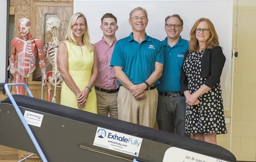
The team consists of people from all disciplines that have come together to pool their knowledge of COPD and patient's pulmonary care and treatment to help these patients to improve their breathing.
The table is based on a gravity-powered approach to improve ventilation as well as helping to clear mucus. The table appears stable but in fact rocks forward and backwards with weight. The person on the exercise table lifts and pulls a bar while rocking the table forward
As the person pushes away the table then rocks backward resulting in the person's feet being higher than their head. This movement forces air out of the lungs, which is normally difficult for a COPD patient to do and therefore reduces the difficulty of breathing for the patient. This approach also uses gravity to help the tiny hairs in the lungs to move the mucus along the trachea as well as the gravity also helping to move the lymphatic fluid out of the lungs. The movement of the abdominal viscera also moves the diaphragm which also reduces the effort of breathing.
The table not only aids the lungs and breathing but also benefits the rest of the body. The gravity effect on the body results in the drainage of lymphatic fluid from the arms and legs, improving circulation and reducing swelling.
One of the founders of the company is himself a COPD sufferer and says that the table has alleviated his symptoms greatly but the table is currently being vigorously tested in trials.
References: http://copdnewstoday.com